Paediatric Non-Accidental Injury: Radiographic Exmaminations

Following on from previous blogs that I have posted, this week I am going to look at Non Accidental Injury (NAI) and the use of radiographic examinations that are carried out within this field.

As stated by the Society and College of Radiographers (2014) within their guidance for radiographers providing forensic radiography services guidelines, all examinations for non-accidental injury are forensic examinations (Society and College of Radiographers, 2014) which are examinations that use application of the science of diagnostic imaging to answer questions of law (Society and College of Radiographers, 2014), as discussed in previous blogs. The Royal College of Paediatrics and Child Health (RCPCH) and the Royal College of Radiologists (RCR) discuss that radiographic imaging of an injured child is critical to the process of child protection (RCPCH & RCR, 2008).

Non- accidental injury is defined as any form of abuse that is purposely inflicted on a person (IMI, 2014), for example emotional, sexual or physical abuse. Sadly it is stated that around 7% of children in the United Kingdom experience serious physical abuse at the hands of parents or carers (Association of Paediatric Radiographers, 2009) with around 55% of children that are fatally abused, being seen within the previous month by a healthcare professional (Association of Paediatric Radiographers, 2009). As radiographers the most common type of abuse that we will come into contact with is physical abuse. It is discussed that radiologists are usually the first professionals to notice signs of NAI (Luijkx & Bhattacharya, 2014), upon the review of radiographs. As set out in the Skeletal Survey for suspected NAI, SIDS and SUDI: Guidance for Radiographers which was set out by the Association of Paediatric Radiographers (Association of Paediatric Radiographers, 2009), when taking NAI into account with children and infants the first radiographic examination should be a skeletal survey conducted by two radiographers with at least one of the radiographers being specially trained (Association of Paediatric Radiographers, 2009). The skeletal survey should include the following plain film projections:

  • AP/Lateral Skull.
  • Lateral Cervical Spine.
  • Lateral Thoracolumbar Spine.
  • Chest X-ray.
  • Left/Right Oblique Ribs.
  • Abdominal X-ray.
  • Left/Right AP Humeri.
  • Left/Right AP Forearm.
  • Left/Right PA Hand.
  • Left/Right AP Femora.
  • Left/Right AP Tibia/Fibula.
  • Left/Right DP Feet (Luijkx & Bhattacharya, 2014).

Where NAI is one of the differential diagnoses a skeletal survey is the standard initial imaging method. Although if the child is suffering from serious life threatening injuries, then a skeletal survey may be postponed, implementing the use of other imaging modalities such as Computed Tomography (CT) (Association of Paediatric Radiographers, 2009). It is important that a skeletal survey is carried out as soon as possible as the outcome may affect the immediate clinical management of the patient and also the management of family/carers, also the early discovery of any suspicious NAI, will result in the child protection team being notified (Association of Paediatric Radiographers, 2009). The resultant radiographs may then be discussed in multi-disciplinary meetings or be used as part of court proceedings in NAI cases (Society and College of Radiographers, 2014).

Under the Ionising Radiation (Medical Exposure) Regulations 2000 either verbal or written consent must be obtained prior to the examination, along with sufficient justification for the examination being provided by the referring clinician (Department of Health, 2000). Whilst the child is in the department along with the two radiographers who are performing the skeletal survey there must be another health professional present who will be responsible for the child’s welfare and safety (Association of Paediatric Radiographers, 2009). The child must be clearly identified, and the identity of the child will need to be confirmed by two radiographers (Association of Paediatric Radiographers, 2009). The skeletal survey should be performed with correct and accurate positioning along with radiographic markers indicating the correct R/L on the images, and to the highest diagnostic quality, the time and date of the exposure should be added as soon as the images are acquired and not at a later date along with the name of the radiographer performing the examinations (Association of Paediatric Radiographers, 2009). It is also very important that the radiographer check previous images.

In conclusion it can be seen that although as radiographers in practice we may not undertake a lot of skeletal surveys for NAI, but the rules and regulations surrounding NAI imaging are vast and must be followed and adhered to. NAI imaging requires at least one specially trained forensic radiographer, although this is quite rare. It is important to mention that the child should be treated with respect, dignity and patience with the radiographer being calm and professional even with the high pressure and seriousness of the situation. It is of the upmost importance that the safety and welfare of the child is maintained to the highest possible standard.


References.

Association of Paediatric Radiographers. (2009, February 1). Skeletal Survey for Suspected NAI, SIDS and SUDI: Guidance for Radiographers. Retrieved November 27, 2014, from Society and College of Radiographers: http://www.sor.org/learning/document-library/skeletal-survey-suspected-nai-sids-and-sudi-guidance-radiographers

Department of Health. (2000). IR(ME)R 2000. London: Department of Health.

IMI. (2014). Non- Accidental Injury. Retrieved November 27, 2014, from Institute of Medical Illistrators: http://www.imi.org.uk/document/non-accidental-injuries

Luijkx, T., & Bhattacharya, B. (2014). Non-Accidental Injury. Retrieved November 27, 2014, from Radiopedia.org: http://radiopaedia.org/articles/non-accidental-injuries

RCPCH & RCR. (2008). Standards for Radiological Investigations of Suspected Non-Accidental Injury. London: The Royal College of Paediatrics and Child Health & Royal College of Radiographers.

Society and College of Radiographers. (2014, May 30). Guidance for Radiographers Providing Forensic Radiography Services. Retrieved November 27, 2014, from SCoR: http://www.sor.org/learning/document-library/guidance-radiographers-providing-forensic-radiography-services/9-non-accidental-injury-nai

A Brief Look At Forensic Radiograpy.

So I have made it to blog 9, and am thoroughly enjoying writing a blog every week – I feel that my writing skills and research skills are developing. For our Diagnostic Imaging cohort last week was classed as a reading week, we did not have any lectures to attend, so for this week’s blog I am going to explore my area of interest – Forensic Radiography. When picking a degree whilst I was undertaking my access course I had to take many things into account – I wanted to pick a career that I would find interesting and would enjoy. Since I have been a little girl I have always had an interest in bones, history and forensics – spending many hours watching the discovery channel and the crime and investigation channel, I have always been fascinated with the human body and what happens after death. I was torn between a career in radiography and a career in forensics and whilst browsing the web one day I came across the term ‘forensic radiographer’ – that was it, I instantly was hooked. I applied to university to study diagnostic imaging – and hopefully one day I will expand on my radiography career and become a forensic radiographer (or that is what I hope!).

Forensic radiography is defined by the Society of Radiographers as the application of the science of diagnostic imaging to answer questions of law, with evidence being collected from either living or deceased subjects (Society of Radiographers, 2010). There are many applications of forensic radiography:

  • Investigation of non fatal injury.
  • Location of forensic evidence.
  • Cause of death.
  • Human identification (Society of Radiographers, 2010).

One particular field of forensic radiography that I am very interested in is the use of non invasive imaging modalities to image ancient remains, or ‘Virtuopsy’ (Virtopsy, 2002-2014), This is where modern imaging techniques are used to perform autopsies – unlike the traditional medical autopsies this technology allows autopsies to be performed without harming the cadaver/remains. Nowadays, with the discovery of ancient remains and remains that have already been discovered that are on display in museums around the world, imaging techniques are being used to examine the remains – this means that they do not have to be damaged or physically examined and that they can be unwrapped virtually. This practice was used within the recent discovery of King Richard the third, when the Kings remains were discovered his body underwent a whole body Computed Tomography (CT) scan as researchers wished to discover the cause of death (University of Leciester, 2014).

Some more information regarding this can be found by clicking the following link:

http://www.defrostingcoldcases.com/king-richard-iii-ruthless-ruler-yes-regicide-no/

CT autopsies are also now being used to give facial reconstructions to discovered remains, such as the ancient Egyptian remains of Tutankhamun. Through the use of Modern CT technology his remains were examined virtually and his face was reconstructed, along with the surprise discovery of many afflictions that he was plagued with (Artnet News, 2014).

Some more information regarding this examination can be discovered by clicking on the following link:

Autopsy Unmasks King Tut’s True Face, and It Isn’t Pretty

Virtual autopsy is also being used in the Trauma patient’s pathway. After death that is deemed as suspicious many patients are now undergoing virtual autopsies so that cause of death can quickly be discovered, along with medical autopsies that are being performed after – the results are found to be accurate and correct. This is supported by a report that documented a case report of three cases of fatal blunt head injury using post-mortem MSCT and MRI which showed massive bone and soft-tissue injuries of the head and signs of high intracranial pressure with herniation of the cerebellar tonsils. Similar findings were found in clinical autopsy which was performed after the digital autopsy (Aghayev, 2004).

Within some cultures, medical autopsies are not allowed for religious reasons – this can mean that families are refusing an autopsy of a deceased patient when it is needed, virtual autopsy is allowed because it is non invasive, this new technology is being seen to overcome cultural and religious barriers (Bakri & Jaudin, 2006).

In conclusion it can be seen that virtual autopsy has a wide variety of uses, and can be used to examine remains in the quickest and most non evasive way.


References

Aghayev, E., 2004. Virtopsy post-mortem multi-slice computed tomography (MSCT) and magnetic resonance imaging (MRI) demonstrating descending tonsillar herniation: comparison to clinical studies. Neuroradiology, 46(7), pp. 559-564.

Artnet News, 2014. Autopsy Unmaskes King Tuts Face. [Online] Available at: http://news.artnet.com/art-world/autopsy-unmaskes-king-tuts-true-face-and-it-isnt-pretty-140493 [Accessed 6 November 2014].

Bakri, D. & Jaudin, R., 2006. Virtual Autopsy, London: Health Technology Assesment Unit Medical Development Division Department of Health.

Society of Radiographers, 2010. Guidance for Radiographers Providing Forensic Radiography Services. 2nd ed. London: Society of Radiographers.

University of Leciester, 2014. Most Likely Cause of King Richards Death. [Online] Available at: http://www2.le.ac.uk/departments/emfpu/Media%20and%20News [Accessed 10 November 2014].

Virtopsy, 2002-2014. About Virtopsy. [Online] Available at: http://www.virtopsy.com/about-virtopsy.html [Accessed 19` November 2014].

Peripherally Inserted Catheter, Central Venous Catheter and Nasogastric Tubes: Mobile x-ray Appraisal Preparation

After our intermediate diagnostic imaging studies practical session this week, which was based on mobile x-rays, and things that need to be taken into consideration by the radiographer before during and after they are called to take a mobile x-ray, I have decided to do my blog this week on the different kinds of lines that radiographers may be required to take an x-ray to check the position of. My blog this week is a descriptive blog, in preparation for my clinical placement in January and also to prepare me for my mobile appraisal, so that if I was called to x-ray to see one of these lines I will know what they are, and also what to look for on an x-ray critique. The lines that I am going to look at in this week’s blog are:

  • Peripherally inserted central catheter (PICC line).
  • Central Venous Catheter (Central Line).
  • Nasogastric intubation (NG tube).

Peripherally Inserted Central Catheter.

A peripherally inserted central catheter is also known as a PICC line, it is a long thin flexible tube (Macmillan, 2014) that is inserted into a peripheral vein (PICC Line Nursing, 2014) that is usually in the upper arm above the elbow. The PICC line is then continually inserted into the vein until the tip of it reaches a large vein close to the heart (PICC Line Nursing, 2014).

Picc 1

(Macmillan, 2014)

A PICC line is inserted by a specialist PICC line Nurse that is trained to insert them, usually the vein in the upper arm is found by the use of ultrasound. After the PICC line is inserted, a chest x-ray is needed to confirm that the PICC line is in the correct place, the patient can either be x-rayed in the department or sometimes a portable x-ray is necessary (Raleighrad, 2014).

picc2N

(Radiology Assistant, 2014)

If the tip of the PICC line is thought to be in the wrong position on the resultant image then it is possible for the specialist nurse to reposition it, and then another chest x-ray is required if again the tip is in the wrong position then the PICC line will be removed and reinserted (PICC Line Nursing, 2014).

A PICC Line is used as an intravenous (IV) line that can administer mediations, such as chemotherapy over a prolonged period of time.

Central Venous Catheter (Central Line).

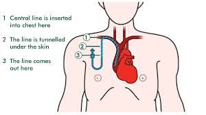
A central venous catheter (central line) is a long flexible tube that is inserted into a vein in the chest or neck, It is then fed into the vein until the tip of the line lies within the proximal  of the superior vena cava (Smith & Nolan, 2013).

cvl1d

(Macmillan, 2014)

The central line can be put in by a specialist nurse or doctor by using ultrasound to find a suitable vein. The patient is either given a local or general anaesthetic. After the central line is inserted a chest x-ray is then needed to check that the tip of the central line is in the correct position (MacMillan, 2014).

cvl2

(Feldman & Gould, 2004)

The central line is used to give intravenous (IV) treatments such as chemotherapy, antibiotics and blood transfusions over a prolonged period of time.

Nasogastric Tubes (NG tubes).

A NG tube is a hollow bored tube that is fed through the nose into the stomach (Patient.co.uk, 2014), and is used for either short or long term nutritional support for the patient.

ng1

(Cancer Research UK, 2014)

After the NG tube has been inserted it needs to have the position of the tip checked, there are two ways of doing this; Firstly a PH test, by using blue litmus paper to check the acidity of the position of the tube – the typical acidity of the stomach is 4, and this should turn the litmus paper blue. Secondly an X-ray of the chest can be used to check the position of the NG tube, the x-ray should be collimated to include the stomach. The x-ray will pick up the position of the tip of the tube if the guide wire is left in (Patient.co.uk, 2014).

ng21

(Patient Safety Authority, 2014)

It is essential that the position of the NG tube is checked, as if it is wrongly inserted it can be fed into the bronchial tree or lung lobe (Patient Safety Authority, 2014), and if a patient is given nutritional support into the bronchial tree/lung lobe instead of the stomach then there can be dire consequences – such as patient death (Patient Safety Authority, 2014).

ng32

(Patient Safety Authority, 2014)


References.

Cancer Research UK, 2014. Cancer Research UK. [Online] Available at: http://www.cancerresearchuk.org/about-cancer/ [Accessed 17 November 2014].

Feldman, J. & Gould, M., 2004. Crossing The Line. [Online] Available at: http://webmm.ahrq.gov/case.aspx?caseID=51 [Accessed 17 November 2014].

MacMillan, 2014. Central Venous Lines. [Online] Available at: http://www.macmillan.org.uk/Cancerinformation/Cancertreatment/Treatmenttypes/Chemotherapy/Linesports/Centrallines.aspx [Accessed 17 November 2014].

Macmillan, 2014. MacMillan Cancer Support. [Online] Available at: http://www.macmillan.org.uk/Cancerinformation/Cancertreatment/Treatmenttypes/Chemotherapy/Linesports/PICCline.aspx [Accessed 19 November 2014].

Patient Safety Authority, 2014. Confirming Feeding Tube Placement. [Online] Available at: http://patientsafetyauthority.org/ADVISORIES/AdvisoryLibrary/2006/Dec3(4)/PublishingImages/23_Fig1.JPG [Accessed 17 November 2014].

Patient.co.uk, 2014. Patient.co.uk. [Online] Available at: http://www.patient.co.uk/doctor/nasogastric-ryles-tubes [Accessed 14 November 2014].

PICC Line Nursing, 2014. PICC Line Nursing. [Online] Available at: http://picclinenursing.com/picc_why.html [Accessed 17 November 2014].

Radiology Assistant, 2014. Radiology Assistant. [Online] Available at: http://www.radiologyassistant.nl/en/p526bd2e468b8c/neonatal-chest-x-ray.html [Accessed 17 November 2014].

Raleighrad, 2014. PICC Line and Port Placement. [Online] Available at: https://www.raleighrad.com/expertise/vein-vascular/picc-lines/ [Accessed 17 November 2014].

Smith, R. & Nolan, J., 2013. Central Venous Lines. British Medical Journal, 65(70), p. 347.

Neonatal Imaging: Use of Imaging Modalities Post Mortem

This week in my Intermediate Diagnostic Imaging studies we looked at neonatal imaging, a lot of neonatal imaging is sadly carried out after death – post mortem, I have decided that for this week’s blog this is a topic that I would like to do a bit of research on and discuss to further my interest in forensic radiography.  Neonatal defines newborn infants being born prematurely or at full term and the care that they receive in the first few weeks of their life (OUP Press, 2011). It has been discovered that sadly 41% of child deaths under the age of five are among new born infants within the first 28 days of their life (World Health Organisation, 2011), with three quarters of neonatal deaths occurring within the first  week of their life (World Health Organisation, 2011). Neonatal death is defined as a death of a child within the first 28 days of life (Patient.co.uk, 2014) and a stillbirth is defined as the death of a baby before or during birth after 24 weeks of pregnancy (SANDS, 2014).

Unfortunately with cases of stillborn or neonatal death the actual cause of death is in a large amount of cases unknown, in cases such as this the foetus or body may be subjected to post mortem imaging to find cause of death, whether it be plain film radiographs, Computed Tomography (CT) scans or Magnetic Resonance Imaging (MRI) or a medical autopsy. The death of a child is devastating to a parent, in a lot of cases in the death of a child the parent wants to know why that child has died. In previous years the child would undergo a post mortem autopsy to discover cause of death, but nowadays with the ever evolving imaging and diagnostic modalities medical autopsies are now being moved away from towards post mortem imaging techniques which are being found to achieve the same results with the correct accuracy and the positive fact that they are non invasive (Bakri & Jaudin, 2006). Usually a post-mortem plain radiograph skeletal survey is carried out with all prenatal and neonatal deaths (Thayyil, et al., 2011), then a conventional autopsy – only with parental consent. A lot of research has been carried out regarding the use of imaging modalities in replacement or adjacent to medical autopsies, although this has mostly been within cases of adult death (Bakri & Jaudin, 2006) the results of imaging modalities used in autopsies were found to be correct and accurate (Aghayev, 2004). Recently, Great Ormond Street Children’s hospital in London have been carrying out research titled ‘Post mortem Magnetic Resonance Imaging in fetuses, newborn and Children: A comparative study with conventional autopsy’ this research was approved by the Great Ormond Street Children’s Hospital research ethics committee (Thayyil, et al., 2011) within this study parents could consent to their child undergoing a MRI and occasionally a CT scan before a medical autopsy would be carried out, the results would be compared to derive the most accurate type of post mortem examination. When considering imaging modalities used within post mortem imaging CT is most commonly used with adults (Department of Health Post Mortem, Forensic and Disaster Imaging Group (PMFDI)., 2012), with prenatal and neonatal imaging MRI is considered the best imaging modality, when carried out alongside plain film radiographs (Department of Health Post Mortem, Forensic and Disaster Imaging Group (PMFDI)., 2012), although if this less invasive post mortem is non conclusive it is advised that the remains be submitted to undergo a medical autopsy alongside it. It is discussed that MRI foetal post-mortem imaging could overcome some weaknesses associated with medical autopsy and furthermore provide a multisystem analysis that is non invasive (Department of Health Post Mortem, Forensic and Disaster Imaging Group (PMFDI)., 2012).

Currently the number of autopsies undertaken within this age group is decreasing rapidly – as parents are not willing to consent to having their child undergo an autopsy as their wish is for their child’s remains to remain intact, this along with refusal of medical autopsy due to religion has lead to this decrease in the number of prenatal and neonatal autopsies being performed. It is discussed that the use of modern imaging modalities in post-mortem ‘virtual’ autopsies could lead to an increase in prenatal and neonatal virtual autopsy numbers (Thayyil, et al., 2011). It is also discussed that autopsies within this age range are essential in the evolution of paediatric medicine and also maternal medicine – to see if there is something that can be done within another pregnancy that could stop the same outcome – the death of a child. (World Health Organisation, 2011).

In conclusion it can be seen that the topic around neonatal are prenatal death is a very delicate one. The ever evolving and progressing field of diagnostic imaging could in the future become a major part of post mortem examinations for this age range, although currently there is still a lot of research that is being carried out around this subject, and its reliability when compared to conventional medical autopsies.


References.

Aghayev, E., 2004. Virtopsy post-mortem multi-slice computed tomography (MSCT) and magnetic resonance imaging (MRI) demonstrating descending tonsillar herniation: comparison to clinical studies. Neuroradiology, 46(7), pp. 559-564.

Bakri, D. & Jaudin, R., 2006. Virtual Autopsy, London: Health Technology Assesment Unit Medical Development Division Department of Health.

Department of Health Post Mortem, Forensic and Disaster Imaging Group (PMFDI)., 2012. Can Cross-Sectional Imaging as an Adjunct and/or Alternative to the Invasive Autopsy be Implemented within the NHS?, London: Nataional Health Service (NHS).

OUP Press, 2011. Oxford English Dictionary. 12th ed. Oxford: OUP Oxford.

Patient.co.uk, 2014. Stillbirth and Neonatal Death. [Online] Available at: http://www.patient.co.uk/doctor/stillbirth-and-neonatal-death [Accessed 15 November 2014].

SANDS, 2014. Stillbirth and Neonatal Death. [Online] Available at: https://www.uk-sands.org/why-babies-die/stillbirth [Accessed 15 November 2014].

Thayyil, S. et al., 2011. Post mortem magnetic resonance imaging in the fetus, infant and child: A comparative study with conventional autopsy (MaRIAS Protocol). BMC Pediatrics, 11(120), pp. 2-11.

World Health Organisation, 2011. Newborn Death and Illness. [Online] Available at: http://www.who.int/pmnch/media/press_materials/fs/fs_newborndealth_illness/en/ [Accessed 15 November 2014].

Stroke Patients: Act F.A.ST

For this week’s blog I am going to look at a patient’s diagnostic pathway, in particular a patient who is thought to have suffered from a stroke. Diagnostic Imaging plays a large part in the diagnosis of a stroke as it is quick and can show a lot of detail for diagnostic purposes quickly. This topic may aid my whilst on placement – possibly as a case study – which would be part of my Intermediate diagnostic imaging studies module, also in the patient health and wellbeing module our groups theme is also stroke, so I could perhaps use this as a reflection within my work.

There are three types of strokes that may affect a patient:

  • An ischemic stroke.
    • This type of stroke occurs when there is an obstruction (clot) within a blood vessel (American Stroke Association, 2014) and occurs in around 87% of stroke patients. (American Stroke Association, 2014)
  •  A haemorrhagic stroke.
    • This type of stroke occurs when a weakened blood vessel ruptures and bleeds into the brain (American Stroke Association, 2014).
  • A transient ischemic attack (TIA)
    • This type of stroke is caused by a temporary blockage (clot) (American Stroke Association, 2014) and are usually warnings of a future Ischemic stroke (Stroke Association, 2014) the symptoms are usually temporary and last for around 24 hours; the blockage either moves or dissolves.

The effects that occur after a stroke can be very debilitating and can sometimes be fatal, depending on the location of the stroke in the brain and how much brain tissue is affected. If the stroke occurs on the left side of the brain – it will be the right side of the body that will be affected along with possible memory loss, slowing down of motor functions and speech and/or language problems. If the stroke occurs on the right side of the brain – it will e the left side of the body that will be affected, along with possible memory loss, vision problems and change of behavioural style. If the stroke occurs within the brain stem – both sides of the body can be simultaneously affected, also stopping motor functions such as speaking (Stroke Association, 2014).

There are certain risk factors within an individual than can increase the likelihood of a patient experiencing a stroke such as, high blood pressure, smokers, diabetic patients, heart disease, age and gender can also impact the likelihood of a stroke, also race and ethnicity (National Heart Lung and Blood Institute, 2014), Symptoms of a stroke tend to develop quickly but can also take hours or days to develop with a TIA the symptoms can last from around 2 to 24 hours (Stroke Association, 2014). Some stroke symptoms include:

  • Sudden weakness.
  • Confusion.
  • Paralysis.
  • Speech/vision difficulty.
  • Breathing difficulty.
  • Loss of consciousness. (National Heart Lung and Blood Institute, 2014)

Within the United Kingdom, there is a lot of campaigning to make the population aware of strokes. The national FAST campaign was introduced to educate the population on strokes and what to look for in an individual.

When stroke strikes, act F.A.S.T

(NHS choices, 2014)

  • F
  • Face: Has their face fallen on one side? Can they smile?
  • A
  • Arms: Can they raise both arms and keep them there?
  • S
  • Speech: Is their speech slurred?
  • T
  • Time: Time to call 999, if you see any single one of these signs. (NHS choices, 2014)

As with the acronym FAST timing is essential when dealing with a stroke patient, a stroke is classed as a medical emergency (The Internet Stroke Centre, 2014).

When a patient is thought to be experiencing a stroke 999 should be dialled immediately, the first medically trained individuals that the patient should occur within the pathway are paramedics. The paramedics will then transport the patient to the emergency department where a doctor should be waiting the patient’s medical history will be obtained along with a physical examination of the patient and blood tests. When a patient is suffering with a suspected stroke there are a multitude of radiographic examinations that may be undertaken:

  • A brain Computed Tomography (CT) scan – this is usually the first diagnostic examination that the patient undergoes as soon as a stroke is suspected, it is quick, very detailed and can show bleeding in the brain or damage to the brain along with other pathologies that may be present resulting in the symptoms the patient is showing.
  • A Magnetic Resonance Imaging (MRI) scan – this can be used additionally to or in place of a CT scan. An MRI scan can detect changes in the tissue of the brain.
  • A CT Arteriogram or an MRI Arteriogram – this test can show the larger blood vessels in the brain and can give information of the location of the blood clot.
  • A Carotid Ultrasound – this examination can show weather the carotid arteries are blocked or narrowed (National Heart Lung and Blood Institute, 2014).

With the use of radiographic diagnostic examinations, patients with suspected strokes can quickly be examined, detailed images of brilliant diagnostic quality can be obtained which can be used to identify if the patient has experienced a stroke, where it is located and what type of stroke has occurred. These images allow a correct diagnosis to be made extremely quickly; this quick diagnosis can allow sometimes life saving medication to be provided to the patient immediately which may improve their quality of life following a stroke.


References.

American Stroke Association, 2014. American Stroke Association. [Online] Available at: http://www.strokeassociation.org/STROKEORG/AboutStroke/TypesofStroke/Types-of-Stroke_UCM_308531_SubHomePage.jsp [Accessed 7 November 2014].

National Heart Lung and Blood Institute, 2014. National Heart Lung and Blood Institute. [Online] Available at: http://www.nhlbi.nih.gov/health/health-topics/topics/stroke/signs.html [Accessed 7 November 2014].

NHS choices, 2014. Stroke: F.A.ST. [Online] Available at: http://www.nhs.uk/actfast/Pages/stroke.aspx [Accessed 7 November 2014].

Stroke Association, 2014. Stroke Associatione. [Online] Available at: http://www.stroke.org.uk/about/types-stroke [Accessed 7 November 2014].

The Internet Stroke Centre, 2014. The Internet Stroke Centre. [Online] Available at: http://www.strokecenter.org/patients/stroke-diagnosis/how-a-stroke-is-diagnosed/ [Accessed 7 November 2014].


Patients with Special Needs: The Elderly

This week for my blog, I am going to look at considerations that need to be taken into account when imaging a patient with special needs (Easton, 2014). This is one of the very first Intermediate Diagnostic Imaging lectures that I attended in my second year; I find the topic extremely interesting as I have a lot of experience when dealing with patients with special needs as I worked as a support worker within this field before attending university. I feel that a lot of the time patients and their needs are not completely understood, so I have decided to do a bit of research around this subject and along with the reflections of my own personal experience produce this week’s blog. It was very thought provoking  to see the range of patients that are classed under the special needs ‘umbrella’, the young, the elderly,  individuals under the influence of drugs and/alcohol, individuals with a disability, learning disorder, individuals with mental health problems or anyone who may need additional aid to undergo their examination. The specific type of patient with special needs that I am going to look at in this week’s blog is the elderly patient.

Whilst in the lecture I discovered that there are four main types of patient’s needs, which are communication needs, physical needs, behavioural needs and cognitive needs. I believe that from my own experience in my career and clinical placement that when encountering elderly patients a lot of the time the certain aspects from one or more of these four of these needs need to be met.

Currently the life expectancy of the population is increasing, it is now stated the within the United Kingdom the average life expectancy age for men is 86 and for females is 89 (Office for National Statistics, 2013). This increasing life expectancy is resulting in individuals developing health problems and being in need of diagnosis, as elderly patients present a wide range of symptoms for numerous conditions it is essential that a correct diagnosis is made so the correct route of care can be decided upon and followed. It is discussed that elderly patients can be distinguished into three categories:

  • Patients in good general health.
  • Vulnerable patients.
  • Frail Patients. (Monfardini & Vecchi, 2013)

Based on these three categories it can be assumed that the treatment of elderly patients in good general health should not really differ much from the treatment of a normal adult patient, although there may be physical needs that may need to be met, for example mobility issues. It can also be assumed that the treatment of vulnerable and frail patients may differ from the treatment of a normal adult patient, as there may be cognitive, behavioural, communication and physical needs that the radiographer may need to comprehend before the patients examination takes place, for example extra staff members to assist with transfers, but then if transfers needed to be made then under the Manual Handling Operation Regulations 1992 (Health and Safety Executive, 1992) all staff involved need to be manual handling trained.

Whilst on clinical placement, at first when encountering individuals with special needs I found it quite difficult when dealing with them, be it knowing how to talk and interact efficiently with them or how to assist them and instruct them without sounding rude. As I got further into my placement I realised quite quickly that if I was polite and tried to understand the patient’s situation before I encountered them then this eased the situation along with showing empathy and patience. Imaging an elderly patient who is classed as vulnerable or frail can be quite challenging especially with the multitude of examinations that a patient can undergo for symptoms that may present. Whilst on clinical placement I encountered a patient who was elderly and required neuro-imaging, this patient was diagnosed with dementia. The patient was to undergo a Computed Tomography (CT) scan of the brain. This CT scan requires the patient to be placed on a bed and be cranially moved into the scanner, the bed is raised quite highly from the floor. This presented to be a challenge as the patient kept trying to get up off the bed, as the individual did not understand the instruction of ‘lie down’. To combat this, the radiographer placed a thin foam pad under the patients head and placed a sheet over the body – to give the impression of lying in bed. Personally I thought that this was a very good technique. Once the patient was lying down and positioned within the scanner the patient was instructed to keep very still, this also presented a problem as the patient could not keep their head still – this presented with movement artefact on the resultant images. It is stated that the problems that a confused patient may present should not preclude the use of imaging or interventional examinations that they may be required to undergo (O’Brien, et al., 2009).

It is important to note that all patients no matter how they present themselves should be treated with dignity and respect as stated by Article 8 of The Human Rights Act 1998 (Equality and Human Rights Commission, 1998). The Society of Radiographers Code of Conduct and ethics also states that Radiographers should demonstrate respect for individual dignity, belief, culture and autonomy through a commitment to the principles of consent and confidentiality (Society of Radiographers, 2008).

It is the role of the radiographer to provide a high standard of care to every patient, also to access the needs of a patient prior to the examination. When challenging patients present the radiographer should be calm and act patiently. Explaining the procedure thoroughly and also what is happening at each step throughout the examination. It is very important that the radiographer completes a three point identification check on each patient as this is a legal requirement under the Ionising Radiation (Medical Exposure) Regulations 2000, this may be hard to obtain with an elderly patient as there may be communication issues – for example the patient may be hard of hearing or may not understand what is being asked of them. In instances like this there may be an identification bangle around the patient’s wrist and there may also be a nurse accompanying them to the department. It is essential that the radiographer checks that this is the correct patient for the correct examination.

It is paramount that every patient be treated as an individual, receiving the best standard of care that is achievable. Good communication skills are an essential part of being a radiographer, this was proved by a study that was carried out in 2007 entitled ‘Effectiveness of Communication between Radiographers and Patients at C W M H Radiology Department, Fiji’ (Kaushal, 2007) where the patients were given questionnaires, interviews were conducted and patient – radiographer consultations were observed, this study showed that in this particular hospital radiographer were lacking in communication skills and patients felt that the standard of care that they received was poor, leading on from this study changes were made and communication training was implemented to improve service user experiences. The Society of Radiographers Code of conduct and ethics discussed communication with elderly patients and they state that good communication with vulnerable people such as the elderly or special needs patients is imperative; Radiographers must always listen carefully to them and respect their views, also being able to identify individuals with communication difficulties and make adjustments to accommodate their particular problems (Society of Radiographers, 2008). I believe that from my own experiences good and effective communication can quickly put a patient at ease, especially with and elderly individual.

In conclusion as mentioned earlier, each patient – regardless of their situation or how they present themselves should be treated with dignity and respect and as an individual. Individuals who are classed as a ‘special needs’ patient are still an individual within their own right – and entitled to the best patient centred care that can be provided to them, even though special considerations may need to be made by the radiographer, adapting techniques and good communication skills are key to tackling patients who may be difficult to image – which as described by the Society of Radiographers is indefinitely the role of a radiographer.


References

Easton, S., 2014. Imaging the Patient with Special Needs. [Sound Recording] (University of the West of England).

Equality and Human Rights Commission, 1998. The Human Rights Act, London: Equality and Human Rights Comission.

Health and Safety Executive, 1992. Manual Handling Operation Regulations. [Online] Available at: http://www.hse.gov.uk/msd/pushpull/regulations.htm [Accessed 21 October 2014].

Kaushal, K., 2007. Effectivness of Communication between Radiographers and Patients at CWMH Radiology Department, Fiji. Biomedical Imaging and Intervention Journal, 3(12), p. 225.

Monfardini, L. & Vecchi, V., 2013. Limitations of Diagnostic Radiology for Frail and Vulnerable Elderly Cancer Patients. Aging Health, 9(3), pp. 283-285.

O’Brien, J., Baerlocher, M. & Myers, A., 2009. Role of Radiology in Geriatric Care. [Online] Available at: http://www.ncbi.nlm.nih.gov/pmc/articles/PMC2628843/ [Accessed 22 October 2014].

Office for National Statistics, 2013. National Life Tables – United Kingdoms, London: Office for National Statistics.

Society of Radiographers, 2008. Code of Conduct and Ethics, London: Society of Radiographers.

Contrast Media: Patient Safety and Adverse Reactions

This week in my Intermediate Diagnostic Imaging Studies module I attended lectures on Pharmacology and Contrast Media (Messer, 2014). Whilst at Clinical Placement this year I was lucky enough to be able to get to watch some CT scans, MRI scans, Procedures that involved Fluoroscopy and Nuclear Medicine scans. Within some examinations of these imaging modalities, contrast media is introduced into the patient’s body; I found it very interesting to be able to witness what this contrast media did to the patient’s body in real time during the examination and the differences the contrast media made to the images acquired but I also got to witness a patient that had an allergic reaction to the contrast media that was introduced into their body for the examination to occur. Leading on from this experience and our lectures I have this week decided to blog about Contrast media and patient safety within Computed Tomography (CT) Scans, specifically iodine based contrast media.

When a patient is due to undergo a CT scan, sometimes it is necessary for the radiographer to administer a contrast media to the patient prior to the examination. Contrast media is a substance that enhances the appearance of certain structures and fluids within the human body (Howell, 2013) and provides diagnostic information that may not otherwise have been attainable. As discussed by the Pharmaceutical Journal (2012) many, if not all contrast media used within imaging modalities is a prescription only medication (Al-Atroshi, 2012), Prescription only medicines are classified under The Medicines Act 1968 and The Prescription Only Medicines (Human Use) Order 1997 (HCPC, 2014), these medications are required to be prescribed by an appropriate practitioner before they can be used or administered.

Within Computed Tomography examinations it is most common that contrast media be introduced into the patient’s body either orally, intravenously or both. Some examinations that require oral contrast media are:

  • Gastrointestinal Tract Examinations such as:
    • Pharynx
    • Oesophagus
    • Stomach
    • Small Intestine
    • Large Intestine (Radiological Society of North America, 2013)

Some examinations that require intravenously administered contrast media are:

  • Internal organs such as:
    • Heart
    • Lungs
    • Liver
    • Adrenal Glands
    • Kidneys
    • Pancreas
    • Gallbladder
    • Spleen
    • Uterus
    • Bladder
  • Gastrointestinal Tract
  • Arteries and Veins
  • Soft Tissues
  • Brain
  • Breast (Radiological Society of North America, 2013)

Although it is thought that contrast media is quite safe to administer to a patient (Radiological Society of North America, 2013) and that less than 1% of the population can have an adverse reaction to the drug (Hunt, et al., 2009) it is still very important to be aware and to make the patient aware of the risks. Whilst on Clinical placement with every examination that I witnessed that involved any type of contrast media the radiographer would ask the patient a series of questions, which I now realise formed part of a strict patient safety questionnaire. The patient was asked firstly, a 3 point Identification check – their name, address and date of birth, this was to ensure that the radiographer had the correct patient for the correct examination and that no mistakes would be made. Secondly, if they had ever had a CT scan? if they had ever been given contrast media before – if so had they had a reaction to it? if they were allergic to iodine? had any kidney problems or were asthmatic. If the patient answered yes to any of these questions, a radiologist would be consulted before the examination and administration of the contrast media took place and it would be up to the radiologist weather the drug would be given. Patient with impaired renal problems should be given special considerations before being administered any iodine based contrast media, as kidney nephropathy can be introduced by iodine.  When the Contrast media is given intravenously a cannula is placed in a vein in the patients hand/wrist by a qualified professional this cannula is then flushed through with saline solution (which is also a Prescription Only Medication) to ensure that the cannula is correctly in situ. The patient is then positioned within the CT scanner and connected to an injection pump which contains the contrast media – which in a lot of examinations is Omnipaque. The patient is then informed that when the injection starts that they may taste a metallic taste, feel a warm sensation and also feel the need to urinate. The radiographer then leaves the room and controls both the scan and the injection via a computer system.

There is always a chance that a patient could have an adverse reaction to any drug, and the reactions are classed into mild, moderate or serious reactions. There can also be immediate or delayed reactions. In all departments that are administering any contrast media, there will be a contrast reaction kit which includes:

  • Epinephrine 1:1000
  • Epinephrine 1:10000
  • Benadryl
  • Solu Cortef
  • Inhaler
  • Extra IV Supplies (Fox, 2014)

Also all radiology departments have designated Nurses, Radiologists, Senior Radiographers and individuals who are Life Support trained. There are also resus trolleys situated in every department.

In my experience of a adverse reaction to a contrast media administered intravenously it was quite a high pressured situation for myself having never witnessed a reaction of that sort before and being a student, it was also a scary situation for the concerned patient who was fine at the time of the injection and had no reaction throughout the examination to then be released from the injection to be brought out of the CT scanner and then start to have quite a severe reaction. Although the radiographer knew exactly how to deal with the situation and immediately called a radiologist and a team of nurses who all rushed to the patient’s aid. Although the patient had been asked all the safety questions and had previously had a CT examination with contrast media and had not experienced a reaction.

In conclusion it can be seen that there are risks associated with administration of contrast media within radiography examinations, but it can also be identified that radiography departments are well equipped and sufficiently trained to deal with any reactions that may occur and also it is worth mentioning that the radiographers who administer the contrast media do ask safety questions, and are very well trained that these reactions can occur.


References

Al-Atroshi, L., 2012. The Pharmaceutical Journal. [Online] Available at: http://www.pharmaceutical-journal.com/opinion/correspondence/contrast-media-are-poms/11106927.article [Accessed 21 October 2014].

Fox, L., 2014. Contrast Reaction Kit Tutorial [Interview] (4 June 2014).

HCPC, 2014. Health and Care Professionals Council – Medicines and Prescribing. [Online] Available at: http://www.hcpc-uk.org.uk/aboutregistration/medicinesandprescribing/ [Accessed 21 October 2014].

Howell, W., 2013. Diagnostic Imaging. [Online] Available at: http://www.diagnosticimaging.com/contrast-agents/communicating-risks-contrast-agents-patients [Accessed 21 October 2014].

Hunt, C., Hartman, R. & Hesley, G., 2009. Frequency and severity of adverse effects of iodinated and gadolinium contrast materials: retrospective review of 456,930 doses. American Journal of Roentgenology, 193(4), pp. 1124-1127.

Messer, S., 2014. Contrast Media. [Sound Recording] (UWE).

Radiological Society of North America, 2013. RadiologyInfo. [Online] Available at: http://www.radiologyinfo.org/en/safety/index.cfm?pg=sfty_contrast [Accessed 21 October 2014].

Trauma Radiography: A Radiographers Role Within A Trauma Team

Recently, in my intermediate diagnostic imaging lectures we have been given two presentations looking at different aspects of trauma within radiography, the role of a radiographer and the radiographer as part of a multidisciplinary trauma team. This along with the fact that trauma radiography interests me and my fascination with the patient pathway of a trauma patient has provoked me to research further into ‘trauma teams’ and the dynamics of a radiographer.

At my previous placement in the Bristol Royal Infirmary I got to witness and participate within a trauma team, I found it exciting but also rather daunting at the same time, as the patient was very unwell and needed immediate care, there were a lot of people with different roles also participating within the patients immediate care needs. It was very interesting to see how all of these separate individuals worked together.

Trauma radiography can be challenging and exciting for a radiographer, however obtaining images of good diagnostic quality can also be extremely stressful. If a radiographer is well prepared, able to maintain composure and also be willing to interact within a multidisciplinary team then this can slightly ease the pressure of the situation.

Trauma is defined as ‘a serious injury or shock to the body, from violence or caused by an accident’. (Farlex. Inc, 2014). Trauma is the fourth leading cause of death in the UK in individuals within their first forty years of life (Sailsbury NHS Foundation Trust, 2013), This is why when dealing with a trauma patient timing is very important, as this could result in the loss of a patient’s life, to minimise the amount of time a radiographer spends acquiring images from a trauma patient, Emergency Departments usually have designated diagnostic equipment located either in the department or extremely close. The first hour after trauma has occurred is called the ‘golden hour’ (Newgard, 2010), and begins the moment the trauma occurs, it is crucial that the patient receives the best possible care within this hour as it can increase the patients chance of survival. The patient while still at the scene of the trauma may encounter paramedics who will transport the patient to hospital where the trauma team will have already assembled and be prepared to deal with any situation that the patient presents.

Once the patient has arrived within the Emergency Department, he/she will encounter a multitude of health professionals; all of these individuals work together and become the ‘Trauma’ team. The trauma team is a multidisciplinary team, within which multiple health professionals work together to promote the best outcome for the patient and to provide the best standard of care. This team consists of:

  • Team Leader
  • Anaesthetist
  • Anaesthetist assistant
  • General Surgeon
  • Orthopaedic Surgeon
  • Emergency Room Physician
  • Nurses
  • Radiographer
  • Scribe

These individuals make up the core of the team, but there may be other members of staff present such as:

  • Porters
  • Haematologists
  • Biochemists

Outside of the immediate trauma team there may also be other health professionals who are indirectly involved within the patients care, such as radiologists, who will authorise additional radiographic procedures that the trauma team leader may require the patient to undergo, theatre staff may also be prepared and waiting if it is thought that the patient may require immediate surgery. Every individual that participates directly or indirectly within the patients’ immediate care needs are all working together as a multidisciplinary team towards the best outcome for the patient, and to provide ‘patient centred care’ which is the main role of the National Health Service (NHS). (Bates & Grimes, 2014).

The role of the radiographer within this team is to start a trauma series of x-rays, firstly starting with a lateral c-spine, then a chest and pelvis x-ray. These views should be obtained in all trauma patients within a trauma situation unless otherwise told by the team leader. (Trauma.org, 2014). It can be hard for the radiographer to safely obtain these images and comply with radiation protection legislations as there may be a lot of individuals within a small immediate area. Radiation Protection is one of the most important duties of the radiographer within a trauma environment; it is the responsibility of the radiographer to protect the patient, trauma team members and them self. In some high risk trauma situations it is impossible for certain members of the trauma team to leave the patient for an x-ray to be obtained, in this instance the radiographer must ensure that team members are wearing lead aprons and that all other team members have moved away to a safe location. (The Royal College of Radiologists and The Society and College of Radiographers, 2012)

The role of a radiographer is described by the Society of radiographers (2003)  as being responsible for providing safe and accurate imaging examinations in a wide range of clinical environments, whilst using a variety of imaging modalities and techniques so that appropriate management and treatment of patients can proceed. I believe that within a trauma situation the role of the radiographer can evolve, it was evident in my experience of a trauma team that not only did the radiographer acquire the image but also had to undertake tasks that may be classed as ‘outside’ a radiographer’s role. Some hospitals specify that a radiographer must answer a trauma call within 5 minutes (The Royal Childrens Hospital, 2012) as the first hour after trauma is referred to as the ‘golden hour’ (Newgard, 2010) and timing is critical. The Radiographer must not leave until they have been dismissed. It is also very important that the radiographer lets the existing team know that they have arrived and also when the images are available to view. There may also be other radiographers involved within this patient’s pathway, for example, if the patient is required to have a CT scan; it is the radiographer’s job to perform this scan.

In conclusion it can be seen that radiography is fundamental within trauma patients’ as it is one of the main diagnostic tools available, and information that may impact on the outcome of a trauma patients care and/or weather anything can be done to help the survive their trauma is gained from radiographic procedures quickly and accurately.

References

Bates, S. & Grimes, K., 2014. Quality Report Kingston NHS, London: s.n.

Farlex. Inc, 2014. The Free Dictionary. [Online] Available at: http://www.thefreedictionary.com/trauma [Accessed 15 October 2014].

Newgard, C., 2010. Emergency Medical Service Intervals and Survival in Trauma: Assesment of the ‘Golden Hour’. Emergency Medicine, 55(4), pp. 235-246.

Sailsbury NHS Foundation Trust, 2013. Sailsbury NHS Foundation Trust. [Online] Available at: http://www.icid.salisbury.nhs.uk/ClinicalManagement/OrthopaedicsAndTrauma/Pages/TraumaTeam.aspx [Accessed 16 October 2014].

The Royal Childrens Hospital, 2012. Trauma Team Compisition Roles. [Online] Available at: http://www.rch.org.au/paed_trauma/guidelines/Trauma_team_composition_roles/#Radiographer [Accessed 16 October 2014].

The Royal College of Radiologists and The Society and College of Radiographers, 2012. Team Working in Clinical Imaging, London: The Royal College of Radiologists and The Society and College of Radiographers.

Trauma.org, 2014. Trauma.org. [Online] Available at: http://www.trauma.org/archive/resus/traumateam.html [Accessed 15 October 2014].

Article Critique: “Pulmonary Embolism in Pregnant Patients: Fetal Radiation Dose with Helical CT”

This week at University I attended a lecture on Radiobiology as part of our Intermediate Diagnostic Imaging studies. The lecture covered the effects of radiation within cells and resultantly the human body as a whole. The lecture also touched on the radiation doses that foetus’ can be exposed to, as part of our blog we have been given an article that is related to this topic to read over and critique.

The article that I am going to critique was written by Helen T. Winer-Muram, MD, in 2002 and is called “Pulmonary Embolism in Pregnant Patients: Fetal Radiation Dose with Helical CT” (Winer-Muram, 2002) it is a medical physics article and its purpose is to calculate mean foetal radiation doses reported by scintigraphy. Scintigraphy is a diagnostic test in which a two-dimensional picture of a body radiation source is obtained through the use of Radioisotopes (Medicinenet, 2014). The aim of this article is to give readers a detailed explanation of the methods of the experiment, how the experiment was ultimately carried out and the results and outcomes that were obtained on completion of the experiment.

This article was published in August 2002 and is now 12 years old; due to its age and with technology developing so rapidly may no longer be viewed as a creditable source. Due to the incredible amount of advancement it CT over the past years since the experiment was carried out, if the experiment were to be undertaken it may not be possible to obtain the same results.

Helen T Winer-Muram, MD is a practising Radiology Doctor with 41 years experience (WebMD, 2014), She specializes in Diagnostic Radiology and Cardiothoratic Radiology (WebMD, 2014). Helen T Winer-Muram, MD has contributed to 71 publications (Vitals, 2014). This makes her a creditable source. Other individuals that contributed to this article are John M. Boone, PhD, who is a professor of radiology (UC Davis Health System, 2014), Haywood L. Brown, MD who is a maternal-fetal medicine specialist (Duke Medicine, 2014), William C. Mabie, MD, who specializes in Obstetrics and Gynaecology, internal medicine and paediatrics (Vitals, 2014), Gerard T. Lombardo, MD who is a pulmonologist (Vitals, 2014) and S. Gregory Jennings, MD. All of these individuals have multiple publications and are all creditable sources adding to the value of this article.

This Experiment was carried out with 23 partcipants, certain test measures/protocols (CT parameters) were kept continuous throughout the study, these were:

  • 120KkVp and 100Ma
  • The scan time: 1second per scintillation
  • Collimation: 2.5mm
  • A pitch of 1
  • Patient position: Craniocaudal
  • Scan extent: 11 cm

With these protocols the mean foetal dose that would be received whilst undergoing a CT scan of the chest was calculated using Monte Carlo techniques. The Monte Carlo technique is a ‘problem solving technique used to approximate the probability of certain outcomes by running multiple trial runs, called simulations, using random variables’ (Palisade, 2014). Also the ‘Monte Carlo simulation is a method for exploring the sensitivity of a complex system by varying parameters within statistical constraints’ (The Mathworks inc, 1994-2014). The article then goes on to explain how the measurements were made and the differences between the cylindrical size that was used and an actual foetal shape and how this would affect the obtained results, ‘Differences in shape between a cylinder and the actual fetus have only a small effect on the fetal dose calculation, as long as the cylinder dimensions simulate the bounds of the fetus’ (Winer-Muram, 2002).

The experiment only used a small sample of pregnant patients, 23 in total, this could have been improved by using more pregnant patients, as ‘Increasing sample size can also give greater power to detect differences’ (Select Statistical Services, 2014) and also it is not specified as to how this sample of patients were picked. These 23 Pregnant Patients were all healthy women, of mixed ages – the mean age was 37 indicating that the patients were of a wide range of ages, mixed body mass indexes and in different stages of pregnancy. Eight of the patients were in the first trimester, nine in the second trimester and six in the third trimester, the results obtained from the experiment would have been more reliable if more pregnant patients were used in each trimester and if all of the patients were of similar age and body mass index.

The materials and methods section of the article was very detailed, it discusses how the experiment was implemented on the phantoms and the doses used.

In the conclusion all of the findings of this research are summarized, showing that helical CT scanning results in a lower foetal dose during all three trimesters.

The article is well presented and has diagrams, although I feel that the results section and the discussion section could have been elaborated on.

In conclusion this article and the data presented within it are relevant to the radiography world and also reinforce dose management and patient safety. There could have been certain improvements that could have been made to make the experiment more creditable and reliable, but it was extremely interesting.

References

Duke Medicine, 2014. Duke Medicine. [Online] Available at: http://www.dukemedicine.org/find-doctors-physicians/#!/haywood-l-brown-md [Accessed 6 October 2014].

Medicinenet, 2014. Medicine Net. [Online] Available at: http://www.medicinenet.com/script/main/art.asp?articlekey=9136 [Accessed 6 October 2014].

Palisade, 2014. Palisade. [Online] Available at: http://www.palisade.com/risk/monte_carlo_simulation.asp [Accessed 6 October 2014].

Select Statistical Services, 2014. Select Statistical Services. [Online] Available at: http://www.select-statistics.co.uk/article/blog-post/the-importance-and-effect-of-sample-size [Accessed 8 October 2014].

The Mathworks inc, 1994-2014. Mathworks. [Online] Available at: 2014 [Accessed 6 October 2014].

UC Davis Health System, 2014. UC Davis Health System. [Online] Available at: http://www.ucdmc.ucdavis.edu/radiology/faculty/boone.html [Accessed 6 October 2014].

Vitals, 2014. Vitals. [Online] Available at: http://www.vitals.com/doctors/Dr_Helen_Winer-Muram/credentials [Accessed 6 October 2014].

WebMD, 2014. WebMD Physician Directory. [Online] Available at: http://doctor.webmd.com/doctor/helen-winer-muram-md-8b6ad87c-92f7-4718-8187-ecb794657eca-overview [Accessed 6 October 2014].

Winer-Muram, H. T., 2002. Pulmonary Embolism in Pregnant Patients:Fetal Radiation Dose with Helical CT. Radiology, 224(2), pp. 487-492.

A Little About Myself……

Hello…… 🙂

My name is Natasha and I am 25 years of age. I am from a town in South Wales called Carmarthen, living in a small little village just outside called Abergwilli. In September 2013 I moved to Bristol, to go to university. Deciding to go to university at the age of 25 and moving away from home and the life I had made for myself since leaving school was a drastic change. Since leaving school at the age of 17 I had worked in various different roles throughout the health sector, starting off as a domiciliary carer, working as a support worker within the adult, children and adolescent mental health teams within Carmarthenshire and finishing up as a support worker within a company called Crossroads, which provided rest bite care to individuals and their families within the community – which allowed service users to remain living at home whilst giving their families a break. I thoroughly enjoyed my role within the Crossroads team. When I was 22, I decided that I wanted to create a better life for myself and decided to go back to college to undertake an access course, which I did part time alongside my job. After completing 1 year of my access course I applied to University to study Diagnostic Imaging, I got an offer from a few universities but decided that I wanted to study at UWE. September 2013 came and I moved to the Glenside campus halls and very nervously started my first day in uni.

1966881_10201199373141079_1108934815_n


This is the September 2013 Cohort, Diagnostic Imaging

Outside of academia, I have a dad, called Chris, a mum, called Gaynor and a (not so) little sister called Laura. Laura also attended UWE to study Diagnostic Imaging and is now a qualified radiographer, she helps my greatly within my course. I have a dog, called Lillie who is a miniature jack Russell and has a very loving/naughty personality and gets up to all sorts of mischief when nobody is watching. I also have a partner called Ben, to whom I recently got engaged to. I am very lucky in the respect that I have so much support in what I am currently doing from family and friends.

     546795_10151532544828517_556963071_n10590481_10201940915239168_2206424243033907614_n10425869_10202124220341681_7235448954000965028_n

I have made lots of friends in uni and also have friends that live in Carmarthen. In Bristol I live with two fellow radiography students, Jessica and Rob and also Liam who is a student paramedic. In my spare time I enjoy socializing with friends, reading, watching films and driving. I have played the piano since a very young age and also enjoy swimming and motor sport. I enjoy going back home at weekends to see my family, friends and also my dog, who is being looked after by my parents whilst I am at university. Whilst at university I have a job as a radiography assistant and I am looking forward to still having an active involvement within the radiography world alongside my studies and placements.

I have an interest in Forensic Radiography and one day I would like to specialize within this field, also having a keen interest in anthropology and forensics.

More information can be found on forensic radiography here:

http://afr.org.uk/about-us/

I have started this blog as part of my second year radiography studies, aiming to learn how to write reflectively and to improve my communications skills.